Ac Joint Disruption Orthobullets

Acromioclavicular Joint Injury Shoulder Elbow Orthobullets

Shoulder Resurfacing Shoulder Elbow Orthobullets

Elbow Physical Exam Shoulder Elbow Orthobullets

Triceps Rupture Shoulder Elbow Orthobullets

Palmaris Brevis Anatomy Orthobullets Com With Images

Clavicle Shaft Fracture Pediatric Pediatrics Orthobullets
Acromioclavicular joint injury 8 22.
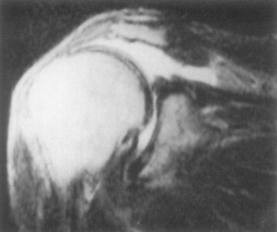
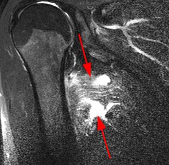
Ac joint disruption orthobullets. Images from his mri are shown in figures b and c. Involves 40 cephalic tilt view of sternum and clavicle. Best assessed radiographically by serendipity view. Fractures and dislocations of the shoulder.
Shoulder resurfacing 10 18 2019 233 views. Clavicle rotates 40 50 posteriorly with shoulder elevation. 8 of rotation through ac joint. Luxatio erecta inferior glenohumeral joint dislocation injuries in throwing athlete slap lesion internal impingement.
Majority of motion is from the bones not through the joint. Diarthrodial saddle joint incongruous 50 contact fibrocartilage not synovial cartilage contains an intra articular disc. A acromioclavicular joint injury otherwise known as a shoulder separation is a traumatic injury to the acromioclavicular ac joint with disruption of the acromioclavicular ligaments and or coracoclavicular cc ligaments. The ac joint is a diarthrodial joint.
Most recently he had another episode of instability when reaching into the back seat while driving. He has weakness performing the physical exam maneuver shown in figure a. Epidemiology demographics more common with age but can occur by second decade of life. Elevation of arm to 90 leads to rotation of the sternoclavicular joint of 30 imaging.
Treatment is immobilzation or surgical reconstruction depending on the degree of separation and ligament injury. Remainder from scapular rotation and sternoclavicular motion. Valgus extension overload pitcher s elbow. Acromioclavicular joint injuries are characterized by damage to the acromioclavicular joint and surrounding structures.
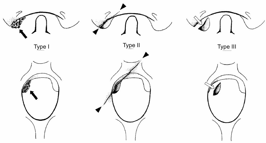
Almost invariably traumatic in etiology they range in severity from a mild sprain to complete disruption. Most common condition of the ac joint ac joint arthritis is caused by transmission of axial large loads through a small contact area resulting in repetitive microtrauma same mechanism as distal clavicle osteolysis. Fibrocartilaginous intraarticular disc is located between the osseous segments. Luxatio erecta inferior glenohumeral joint dislocation.
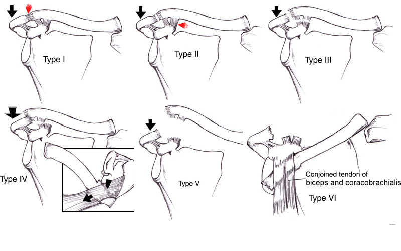
Rockwood classification of acromioclavicular joint separations clinical orthopaedics and related research 475 1. Acromioclavicular joint ac separations are one of the most common injuries seen in orthopedic and sports medicine practices accounting for 9 of all injuries to the shoulder girdle 1 3 various operative and nonoperative treatment schemes have been described for the management of ac joint injuries 4 33 although consider.
Occipital Condyle Fractures Spine Orthobullets
Latissimus Dorsi Rupture Shoulder Elbow Orthobullets

Humeral Avulsion Glenohumeral Ligament Hagl Shoulder Elbow

Shoulder Instability Orthobullets Orthopedic With Purcell At
Deltoid Rupture Shoulder Elbow Orthobullets

Shoulder Periprosthetic Fracture Shoulder Elbow Orthobullets

Unsable Slipped Capital Femoral Epiphysis Emergency Department
Ortho Blog Cmc Compendium

Proximal Humerus Anatomy Met Afbeeldingen

Long Major Bones Tibia General View Medial Condyle Lateral

Pin On School

Scapulothoracic Dissociation Dr James Mclean
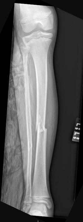
Tibial Shaft Fractures Pediatric Pediatrics Orthobullets
