Ac Joint X Ray Grade 2

Acromioclavicular Joint Injury Type 3 Radiology Case Radiopaedia Org

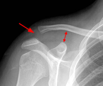
Rockwood Classification Of Acromioclavicular Joint Injury Annotated Radiographs Radiology Case Radiopaedia Org

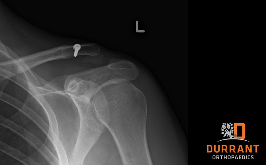
A Anteroposterior X Ray Of A Left Shoulder In Which An Acj Injury Download Scientific Diagram
Rockwood Classification Of Acromioclavicular Joint Injury Radiology Reference Article Radiopaedia Org

Xwfflmzk 3gcxm

Ac Joint Separation Radiology Acromioclavicular Joint Injuries Usually Occur From A Direct Blow Or Following A Fall Onto The Shoulder With An Adducted Arm Thi
X ray and mri finding.
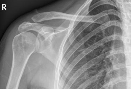
Ac joint x ray grade 2. The medical term for this is subluxed. Localised pain swelling and deformity. Jeff padalecki orthopedic shoulder specialist in the greater austin texas area. No deformity visible clinically or on x ray grade 2.
Tossy classification tossy et al corr 28. Low grade ligament injury may not be visible on a plain x ray the acromioclavicular joint can be assessed with standard shoulder x rays. The joint is incompletely dislocated. Comparing the injured side to the normal side one can see an asymmetry when examining the space between the clavicle and coracoid processs coracoclavicular interval.
For more resources on the ac joint separation grading scale or to determine your grade of injury with the assistance of an ac joint separation grading x ray and mri please contact dr. This well known 6 type system is a modification of the earlier 3 class classification system described by allman 1967 2 and tossy 1963. Involves inferior displacement of the distal clavicle either subacromial or subcoracoid. Acromioclavicular joint dislocation grade 2 3.
Almost invariably traumatic in etiology they range in severity from a mild sprain to complete disruption. The original grading system had three grades. Non displaced sprain type 1 partially dislocated joint type 2 and completely dislocated type 3. Two views of right shoulder show elevation of the clavicle and separation of the ac joint in a type iii ac joint separation.
Interpreting x rays of the shoulder joint duration. A grade 2 ac joint separation results from an incomplete tearing of the acomioclavciular and or the coracoclavicular ligaments. It takes into account not only the acromioclavicular joint itself but also the coracoclavicular ligament the deltoid and trapezius. Strain and contusions of ac joint.
The rockwood classification 1998 is the most common classification system in use for acromioclavicular joint injuries 3. 111 119 1963 grade 1. Breaking down the differences in ac joint sprains duration. Like many orthopedic conditions ac joint separations can range from mild to very severe.
On x ray there will be a 2 3 times increase in the distance between the coracoid and clavicle or a 100 300 increase in the clavicle acromion distance. Fracture of distal end of clavicle is frequently associated with cc tears with or without separation of ac ligament. X rays show one half separation of the ac joint ie clavicle displaced cephalad by one half the depth of the ac joint. Acromioclavicular joint injuries are characterized by damage to the acromioclavicular joint and surrounding structures.

Shoulder Dislocation Radiology Reference Article Radiopaedia Org
Ac Joint Seperations And Injuries A Patients Guide

Pulmonary Hamartoma Radiology Case Radiopaedia Org Radiology Hiatus Hernia Pulmonary

Acromioclavicular Joint Ap View Radiology Reference Article Radiopaedia Org

Ac Joint Separations Overview Of Diagnosis And Treatment

Shoulder External Rotation View Radiology Reference Article Radiopaedia Org

Os Radiostyloideum And Triquetral Fracture Radiology Case Radiopaedia Org With Images Radiology Radiologic Technology Radiology Technologist

Separated Shoulder Wikipedia

Acromiohumeral Interval Radiology Reference Article Radiopaedia Org

Pin On Oh The Joys Of The Medical Field

Supraspinatus Outlet View Shoulder Xray Demonstrates Outlet Impingement Of The Supraspinatus And Coracoacromial Arch With Images X Ray Shoulder Outlet

Pin Di Radiology Intersting Cases